Research

Our laboratory is focused on understanding the allosteric mechanisms that allow macromolecular complexes to perform and regulate their function.

Cryo-electron microscopy gives us the unique opportunity to obtain experimental molecular movies to depict these macromolecules in action, a paramount goal of structural biology. We aim to use this unique opportunity to shed light on pharmacologically-relevant processes with a particular focus on mechanisms happening at the cell membrane.

Signaling at the Membrane

Many signaling pathways rely on the transmission of signal across cell membranes. Our laboratory is focused on understanding the allosteric mechanisms that allow transmembrane proteins to couple ligand binding on one side of a membrane to signaling events on the other side. These mechanisms are particularly relevant to medecine, as many pharmacological drugs taget those proteins. A better understanding of those allosteric mechanisms would allow us to design more selective and safer drugs, an ultimate goal of our research. 

Ryanodine receptors

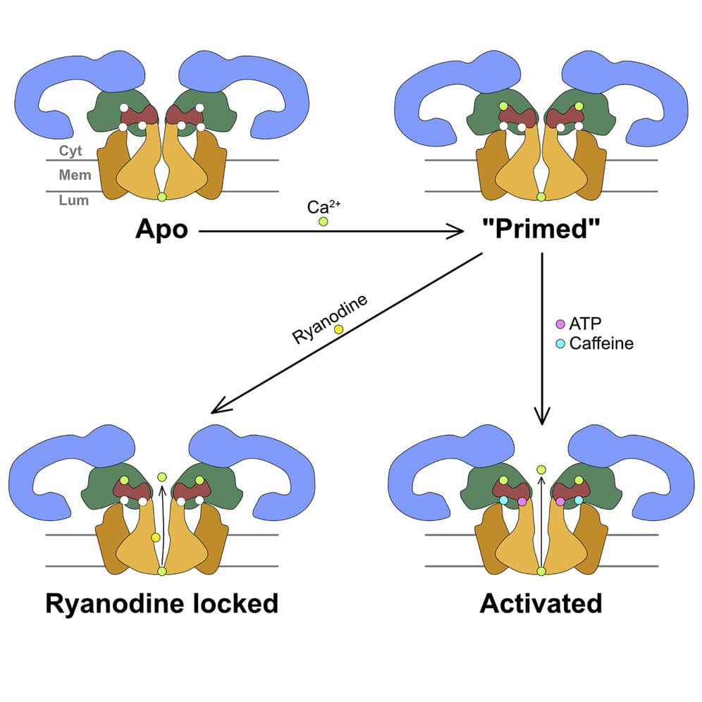
Ryanodine receptors (RyRs) are homotetrameric ~2.2 MDa calcium release channels located on sarco/endoplasmic reticulum membrane (SR/ER). Two isoforms, RyR1 and RyR2, are key elements in skeletal and cardiac muscle excitation-contraction coupling (E-C coupling) that converts electrical signals and rising Ca2+ levels into mechanical output (muscle contraction). E-C coupling controls muscle contraction by the tightly regulated release of Ca2+ from SR stores by RyR channels. Abnormal RyR gating is responsible for muscle disorders including cardiac arrhythmias, heart failure, central core disease and malignant hyperthermia. The molecular mechanism of ryanodine receptors gating and of its refined modulation by small molecule and protein ligands are still not fully understood. We are using single-particle cryo-electron microscopy in conjunction with machine learning image analysis methods to probe the gating mechanism of ryanodine receptors in unprecedented detail. In parallel, we are using cryo-electron tomography to visualize the ryanodine receptors supramolecular assemblies in native membranes. We aim to understand the structural interactions that occur during ryanodine receptors clustering, how clustering affects their function, and how that may dictate the shape of the Ca+ pulse during E-C coupling and in other excitatory cells.

G protein-coupled receptors

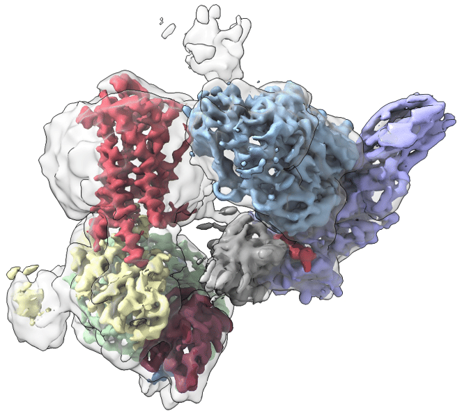
G protein-coupled receptors (GPCRs), also alternatively referred to as seven transmembrane receptors (7TMRs), are the largest class of cell surface receptors and are involved in the regulation of many physiological processes. Their ubiquity in physiology and disease as well as their diversity in structure and function make them very attractive targets for therapeutic development. Currently, it is estimated that over one third of all approved therapeutics target GPCRs.
We aim to take advantage of this golden age that cryo-electron microscopy is bringing to the structural study of GPCRs. Cryo-electron microscopy allows us to study ligand states previously inaccessible and to peer into their dynamics, directly retrieved from the electron microscopy data. This will be particularly useful for structure-based drug discovery efforts and for the development of receptor subtype-specific drugs or biased ligands for the treatment of a variety of diseases.

Host-Pathogen interactions

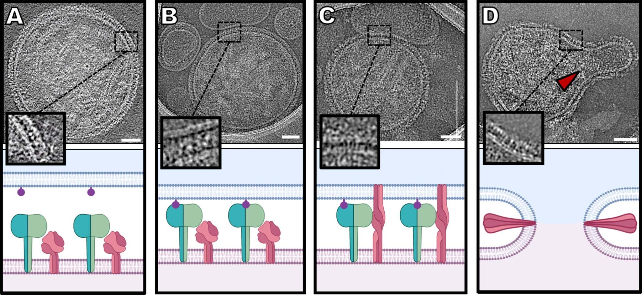
The recent pandemics, including the current COVID-19 pandemic, are showing us how global warming and other changes in our environment are facilitating the transfer of new diseases to human populations, with devastating consequences. At the heart of this problem is the adaptation of pathogens to interact with new hosts. Uncovering the molecular mechanisms underlying these host-pathogen interactions are integral in developing new strategies to prevent or cure infectious diseases. We aim to uncover the dynamic molecular events from initial interaction to entry of pathogens into their host with a combination of single-particle and tomographic cryo-electron microscopy. By deciphering every step leading to pathogen entry, we aim to discover key interactions that will further our fundamental understanding of host-pathogen interactions and help develop more potent inhibitors of the process. Our major focus is on respiratory viruses such as Human parainfluenza virus type 3 (HPIV3) and COVID-19.

Developing methods to study allostery and dynamics in large macromolecular complexes

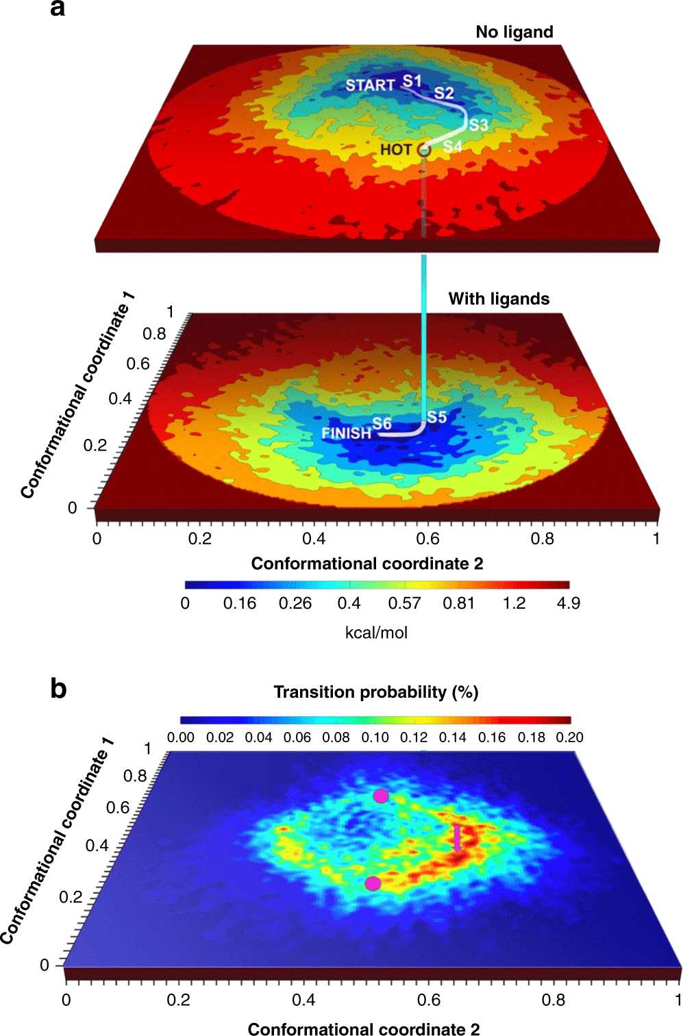
One of our main goals is to be able to decipher long-range allosteric networks in large macromolecular complexes. Physical models cannot yet predict large conformational changes of biological macromolecules even of small size. Such models are key to a true understanding of molecular function and to the predictive design of allosteric drugs. We aim to develop methods that combine current physical models  and experimental ensembles of structures obtained from cryo-electron microscopy data to decipher the allosteric networks that are key to the function and regulation of macromolecular complexes. Towards this goal, we use the ryanodine receptor and the ribosome as model systems where long range allosteric regulation is critical for function. We use machine learning algorithms, such as manifold embedding, to uncover the energy landscape of those macromolecules and how they can be affected .

Manifold embedding

Cryo-electron microscopy gives us the unique opportunity to obtain experimental molecular movies to depict these macromolecules in action, a paramount goal of structural biology. Electron microscopy images capture all the conformations a population macromolecule can assume in solution prior to being snap-frozen on the electron microscopy grid. It gives us the opportunity to harness that information about their conformational dynamics and energy landscape. To shed light on the inner workings of our macromolecules of interest, we are collaborating with the group of Abbas Ourmazd who is developing geometric machine learning algorithms based on manifold embedding that retrieve the energy landscape of our macromolecules and uncover their complex motions. Our lab uses that information combined with modeling and molecular dynamics methods to decipher the complex allosteric networks that regulate our macromolecules’ function.

Translation Initiation

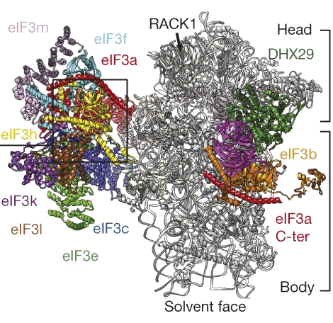
The ribosome has been a major model system for the development of single-particle EM methods.  As a very large RNA-protein macromolecule at the center of protein production and homeostasis, the ribosome is a dynamic brownian machine with complex allosteric regulation. Our lab focuses on understanding the dynamics of the translation initiation step, which is a major checkpoint in the translation cycle. Translation initiation involves many initiation factors, all of which act allosterically on the ribosome to affect the choice of mRNA to be translated, its scanning and the recognition of the start codon. We aim to shed light on how the initiation factors affect the dynamics of these processes in order to better understand how translation initiation is controlled to fine-tune protein homeostasis and the response to environmental stimuli.